Mast Cell Tumors (Mastocytomas) in Dogs
Written by Small Door's medical experts
While some may be benign, mast cell tumors are the most common malignant skin tumors found in dogs and account for 16-21% of all skin tumors in canines. The treatment and prognosis depend on the grade and stage of the tumor.
In This Article
What is a mast cell?
Mast cells are an important part of a dog’s immune system. They play a vital role in the defense against parasite infestations, tissue repair, and the formation of new blood vessels, as well as allergic responses, non-allergic skin disease, wound healing, and tissue remodeling. It is only when these cells replicate in abnormal numbers that tumors can occur.
What are mast cell tumors in dogs?
Mast cell tumors (MCTs) occur when the mast cells (located primarily in the skin, respiratory, and digestive tract) replicate in higher than normal numbers.
Mast cell tumors vary in shape, size, and appearance. They primarily present as a single mass but multiple masses can develop. Your veterinarian will perform a number of tests to determine the extent of the disease. Prognosis is dependent on multiple factors, including the location, grade, and treatment given.
Signs & symptoms of mast cell tumors in dogs
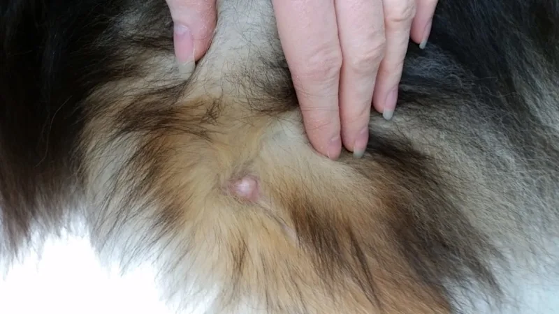
MCTs that present as raised lumps on the skin are often discovered by the dog’s owner. The lump itself may vary in appearance, from a wart-like mass to a soft lump located just beneath the skin or an ulcerated skin mass. MCTs can also be mistaken for insect bites or an allergic reaction.
Tumors located underneath the skin appear as soft nodules and can be misdiagnosed as lipoma, another type of canine skin tumor.
About 50% of all mast cell tumors are located on the trunk and perineum of the body; 40% are found on extremities, such as the paws; and 10% are found on the head and neck region.
Depending on the location and grade of the tumor, an array of symptoms may present themselves:
Fluctuation in size
Rapid growth after months of slow growth or inactivity
Redness and fluid build-up (most common with high-grade skin and subcutaneous tumors)
Enlarged lymph nodes near the tumor
Itchiness and inflammation of the mass (due to higher levels of histamine in the tumor)
Enlarged liver and spleen (when mast cell cancer is widespread)
Loss of appetite, vomiting, and/or diarrhea, depending on the stage of the disease.
What causes mast cell tumors in dogs?
Currently, the underlying cause of MCTs in dogs is unknown, as with many other cancers
Though they are commonly found in older dogs, usually around eight years of age, even puppies as young as a few months old can develop MCTs. Some breeds are more susceptible than others:
Boxer
Boston Terrier
Bulldog
Pitbull Terrier
Weimaraner
Rhodesian Ridgeback
Although these breeds are more vulnerable, it should be noted that MCTs can occur in any dog (and even occasionally in cats).
Grading of mast cell tumors in dogs
Grading refers to a description of a tumor, designed to predict how it will behave, such as whether it will remain localized or spread throughout the body. Once the tumor is biopsied, the pathologist can determine the grade of the tumor based on location, presence of inflammation, and how well the cells are differentiated (i.e. how much a particular tumor cell looks like a normal cell). The more differentiated the cells are, the better the prognosis.
Grade 1 – Cells are well-differentiated. 25% recurrence rate. Low chance of metastasis.
Grade 2 – Moderately differentiated. 44% recurrence rate. Potential for locally invasive metastasis.
Grade 3 – Poorly differentiated. 76% recurrence rate. High potential for metastasis.
Additionally, the pathologist will examine the MCT’s mitotic index, which assesses how quickly the cancerous cells are multiplying. Mast cell tumors with a low mitotic index have a better prognosis than those with a high index.
Stages of mast cell tumors in dogs
The stage of the tumor refers to how much it has spread. Symptoms of MCT may vary depending on the stage of the disease:
Stage I – Single tumor without metastasis.
Stage II – Single tumor with metastasis into the surrounding lymph nodes causing secondary growth.
Stage III – Multiple skin tumors, or a large tumor that has invaded the subcutaneous tissues. There may or may not be lymph node involvement.
Stage IV – Presence of one or more tumors with metastasis in the skin and possibly other organs. Lymph nodes will be involved.
Diagnosing mast cell tumors in dogs
Fine Needle Aspiration
Mast cell tumors are typically diagnosed using fine needle aspiration. This process involves taking a small needle with a syringe and suctioning out a sample of cells straight from the tumor and putting them on a microscope slide that is examined by a veterinary pathologist.
Biopsy
Another method used is called a biopsy. Biopsies, or surgical tissue samples, can be extremely beneficial for situations in which monitoring the aggressiveness of a tumor is critical for managing the growth — this is especially true for MCTs.
Mast cell tumors are notorious for resembling other skin reactions or growths, such as insect bites, warts, and other types of skin tumors. That’s why it’s important to contact your veterinarian if you notice any skin abnormalities.
Once a diagnosis of mast cell tumor has been made, a veterinarian might recommend a prognostic panel on a tissue sample from the tumor so they can determine the abnormalities and genetic makeup of the tumor. This information can then be used by the veterinarian to give a prognosis for your dog.
Treatment for mast cell tumors in dogs
Treatment options for MCTs include surgery, chemotherapy and/or radiation therapy. Prognosis and survival rate are dependent on the grade and stage of the tumor, and how soon appropriate treatment is given.
The first step is to remove the tumor, if possible. If an aggressive surgery is performed early on, there is a greater chance for a positive prognosis. For single MCTs, a procedure called a “wide resection” completely removes the tumor and the surrounding tissues. The removed margins are studied by a pathologist to determine whether there are any lingering malignant cells. If the tests come back negative, your doctor will indicate “clean margins.” If the presence of mast cells are found in the remaining tissues of the surgery site, your doctor will indicate “dirty margins,” which will require follow-up treatment.
A Grade I or II tumor that has been completely removed usually does not require any immediate post-operative treatment. A Grade III tumor, multiple tumors, recurrent tumors, or tumors with “dirty margins” generally require follow-up treatment.
If a dog diagnosed with a Grade I tumor does not experience any recurrence 30 weeks post-surgery, he is considered cured. On the other end of the scale, only 10% of dogs diagnosed with a Grade III malignant MCT live more than a year after surgery.
If the primary tumor and/or lymph nodes affected by the disease cannot be taken out completely, chemotherapy might be an option for providing some short-term relief.
If the tumor cells have spread to other parts of the body, removal of the tumor and affected lymph nodes are of minimal benefit. Chemotherapy may provide short-term benefits. Radiation therapy is another option for MCTs located in areas not suitable for surgery.
Side effects of treatment include nausea, vomiting, lethargy and a decreased appetite. Check with your vet for supportive medications to assist if any of these or other side effects develop.
Is there a cure for mast cell tumors in dogs?
If the MCT has a low grade and a low mitotic index, surgical removal of the tumor may result in a cure. Close monitoring is extremely important, especially for those with a history of MCTs, as these dogs may be prone to developing new (not metastatic) tumors.
Are mast cell tumors in dogs contagious for humans or other pets?
MCTs are not contagious for other pets or humans.
What is the cost of treating mast cell tumors in dogs?
The cost to treat MCTs depends on the expense of various factors:
Initial visit
Type and stage of tumor
Surgery
Number of chemotherapy and/or radiation treatments (if necessary)
Follow-up appointments and testing
The cost to remove a tumor surgically can run well over $1000. If chemotherapy and/or radiation is needed, cost is determined by the number of rounds, size of the dog, and the medications used. A veterinary oncologist will also have a higher fee than a regular vet.
Costs will also vary based on where you live. Pet care in larger cities with higher costs of living will often run higher than in other locations.
Recovery and management of mast cell tumors in dogs
A dog’s recovery is dependent on the grade, mitotic index, and location of the tumor, whether it was completely removed, presence of metastasis, and the type of treatment given.
A healthy diet is essential during this time, especially for dogs receiving chemotherapy. Chemotherapy drugs can lower the body’s immunity, so it’s important to protect your dog from other illnesses and communicable diseases during treatment. Proper nutrition also helps maintain a dog’s strength and improves overall response to the treatment itself.
Is there a vaccine for mast cell tumors in dogs?
Currently, there are no vaccines that can be given to prevent MCTs. The best defense is a combination of good health practices and early detection.
Summary of mast cell tumors in dogs
Mast Cell Tumors are the most common malignant skin tumors in dogs, and they present as raised lumps on the skin. Treatment and survival rates vary depending on the stage and grade of the tumor. Early detection and good overall health are the best ways to combat MCTs. Check your dog regularly and consult your veterinarian straight away if you find any mysterious lumps or nodules.
Related articles
 Medical
Diarrhea in Dogs
Diarrhea in dogs—commonly defined as loose, watery, or runny stool—has a wide range of possible causes. Some of the diseases or conditions that produce diarrhea are mild, while others are more serious.
 Medical
Vomiting in Dogs
As unpleasant and distressing as it might be, vomiting is not uncommon for dogs. There are numerous contributing factors that can lead to this uncomfortable yet important function. If a dog throws up once and is otherwise behaving normally, this generally isn’t cause for concern. However, vomiting can be a sign of a more serious health issue, particularly if other abnormalities – such as diarrhea, lethargy, or loss of appetite – are present. Additionally, young puppies that haven’t yet been fully vaccinated are at greater risk of contracting a serious disease or infection, so if your puppy is vomiting and you suspect a serious health issue could be the cause, contact your vet immediately.